
At the age of 18 she continued her love of the sciences by studying chemistry at the University of Oxford in England to become a prominent British chemist.
At the age of 28, in December, 1938, shortly after the birth of her child, she was diagnosed with rheumatoid arthritis which would become progressively worse and crippling over time with deformities in both her hands and feet. |
Dorothy Mary Crowfoot Hodgkin
|
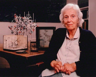
She advanced X-ray crystallography, protein crystallography, and confirmed the structures of cholesterol (1937), penicillin (1945) and vitamin B-12 (1954). It was her life's work to determine the three-dimensional structures of many biologically important molecules using the relatively new technique of X-ray diffraction. For this pioneering work, she was awarded the Nobel Prize for Chemistry in 1964. (1, 2, 3)
|

|
People with Methylmalonic Acidemia (MMA) and cobalamin disorders (B12) may have difficulty with growth and development, neurological problems such as strokes, seizures and low muscle tone, kidney problems, poor vision, and metabolic instability causing them to become seriously ill, sometimes with little warning. There is no cure for any type of MMA, but special diets, vitamin therapies and in some patients, organ transplantation, are used for treatment.(1)
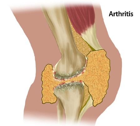
Untreated Bleeding in the Joints > Complications > HoG Handbook > Hemophilia of Georgia https://www.hog.org/handbook/section/4/untreated-bleeding-in-the-joints Blood flowing into the space in the synovial joint causes damage to the joint. If this happens often enough, it causes crippling arthritis. The person will not be able to use the joint without a lot of pain. To show how this happens, we will look at a normal knee joint and compare it to one that has bled over and over. |
|
The joint and bones are slowly destroyed. The cartilage continues to erode away. The unprotected ends of the bones begin to spread out. As the ends of the bones shift, they rub together. This causes intense pain when you move. Bending the knee becomes almost impossible since the joint contains so much scar tissue.The joint wastes away. What is left is a painful condition called degenerative arthritis. The knee is too stiff and painful to move easily. The muscles become weak from not being used. This makes them more likely to bleed.
It is a vicious cycle. When a joint bleed is not quickly or completely treated, the joints and bones are damaged. The muscles weaken, then more bleeding occurs. The end result is crippling. If this happens, physical therapy or surgery may help.
Read Full Article: Untreated Bleeding in the Joints > Complications > HoG Handbook > Hemophilia of Georgia
https://www.hog.org/handbook/section/4/untreated-bleeding-in-the-joints
Food, Blood and Bones By Denise Walker
books.google.ca/books?hl=en&lr=&id=EwLzKMqoIVYC&oi=fnd&pg=PA2&dq=blood+gets+thicker+you+get+joint+pain.&ots=7tomzs4g-I&sig=fEE0VOI_6RxTPl7VdOLDUqOZ0FE&redir_esc=y#v=onepage&q&f=false
|
Central Nervous System Vasculitis (CNS Vasculitis)
Central Nervous System (CNS) vasculitis is an inflammatory brain disease targeting the blood vessels of the brain and/or spinal cord. In this disease, cells of the immune system attack the brain blood vessel walls, which leads to swelling and damage of the wall itself and the surrounding brain tissue. Sometimes, part of the artery wall becomes inflamed, which can lead to swelling of the wall. This expansion or swelling of the artery wall can secondarily narrow the lumen of the artery, through which blood flows into the brain. If the narrowing is severe enough, there may not be enough blood flow to the brain and the patient may start to experience symptoms of stroke. Finally, the artery can close altogether. What labs should be included in the work up of stroke/concerns for cPACNS?
https://www.hindawi.com/journals/crin/2014/868590/ |
There are many different kinds of vasculidites (inflammations of vessels). Most cases of CNS vasculitis occur as a part of autoimmune or inflammatory disorders, such as:
Symptoms include:
1. severe headache, long-lasting 2. strokes (transient ischemic attacks) 3. forgetfulness/confusion 4. weakness 5. vision problems 6. seizures 7. encephalopathy 8. sensation abnormalities https://www.facebook.com/ExemplifyHealth/videos/5991291050978422
How the body handles the COVID VAX when having Type 2 Diabetes - "The Breakthrough Infection" https://www.facebook.com/watch/ExemplifyHealth/what are small amounts of
|
|
|
Giant Cell Arteritis
You know the patient who comes in with cranial pain, scalp tenderness, has some polymyalgia type of symptoms or an octogenarian who's lost 18 pounds and has an anemia of eight point seven grams and a sed rate of sixty, has had colonoscopies ...see Giant Cell Arteritis |
|
|
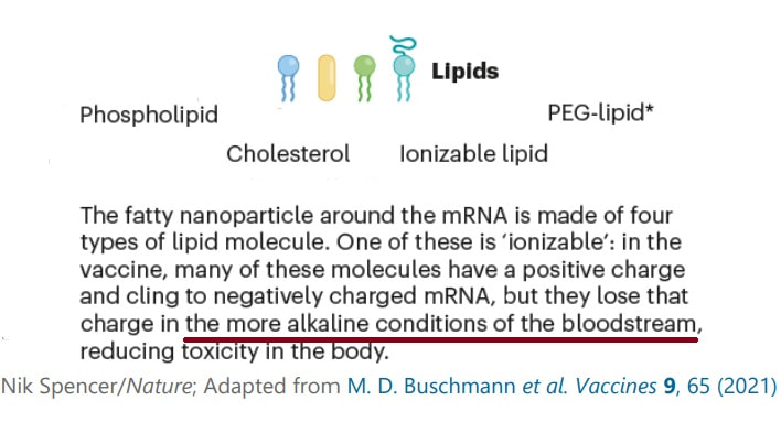
Alkaline vs Acidic body – How to Know If You Are Too Alkaline or Too Acid? – Dr.Berg - https://www.youtube.com/watch?v=m_AKdZ2E1oo |
|
|
|
|
Respiratory acidosis
Respiratory acidosis occurs when too much CO2 builds up in the body. Normally, the lungs remove CO2 while you breathe. However, sometimes your body can’t get rid of enough CO2. This may happen due to:
|
Metabolic acidosis
Metabolic acidosis starts in the kidneys instead of the lungs. It occurs when they can’t eliminate enough acid or when they get rid of too much base. There are three major forms of metabolic acidosis:
the urine pH will be greater than 5.3 or to 5.5 Acid-base balance » |
|
Anemia
|
|
- Some types of anemia are caused by a deficiency or dysfunction of erythropoietin, a hormone produced by the kidneys that stimulates the bone marrow to produce RBCs. A lack of erythropoietin can lead to decreased red cell production by the bone marrow.
|
Can Vitamin B12 deficiency cause arthritis?
Too little vitamin B-12 can cause exhaustion, cognitive difficulties, nerve damage and anemia. B12 is bound to animal protein, its activity is retained during the cooking of most foods. B12 must be released from this protein by pepsin - a digestive enzyme in the stomach.
Paragraph. ここをクリックして編集する.
|
Folate, Folic acid (Vitamin B9)
Can RA cause low iron?
|
Iron Toxicity Post #75: PROOF that Copper Deficiency is the CAUSE of so-called ‘anemia’ therootcauseprotocol.com/iron-toxicity-post-75-formerly-itp76/
- Copper Toxicity and How to Reduce Elevated Levels www.holistichelp.net/blog/copper-toxicity-and-how-to-reduce-elevated-levels/
Do you suffer from any of these conditions?
Chronic fatigue, fibromyalgia, Lyme disease, Hashimoto’s, rheumatoid arthritis, multiple sclerosis, polycystic ovary syndrome…
The Root Cause Protocol. is all about repairing cellular dysfunction to resolve various autoimmune conditions including, but not limited to, the list above. Even though conditions and symptoms will vary from person to person, the fundamental root cause of how they started is the exact same for everyone.
|
Phase 1FOUNDATIONS
1. START Taking adrenal cocktails 2. START Taking mineral drops or applying transdermal magnesium 3. START Taking wholefood vitamin C (WFC) complex 4. START Taking magnesium 5. START Eating grass-fed organic beef liver Phase 2SUPPORTING NUTRIENTS 6. START Eating organic ancestral whole foods and drinking mineralized filtered water 7. START Taking Mother Nature’s sources for B vitamins 8. START Taking wholefood vitamin E complex 9. START Taking boron 10. START Taking cod liver oil |
Phase 3ADVANCED NUTRIENTS
11. START Taking taurine 12. START Taking silica / diatomaceous earth 13. START Taking iodine from food Phase XDEEPER SUPPORT 14. START Donating blood 15. START Managing histamine levels/reactions 16. START Releasing emotional stress 17. START Strengthening the bioenergetic field 18. START Getting regular sunlight 19. START Doing joyful movement Source: RCP 101 Video Series | The Root Cause Protocol https://therootcauseprotocol.com/rcp-101-video-series/ |
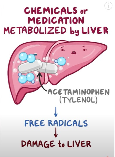
Free radical damage - causes, symptoms, diagnosis, treatment, pathology
https://www.youtube.com/watch?v=2ZRAUO51Wf8
https://www.youtube.com/watch?v=2ZRAUO51Wf8
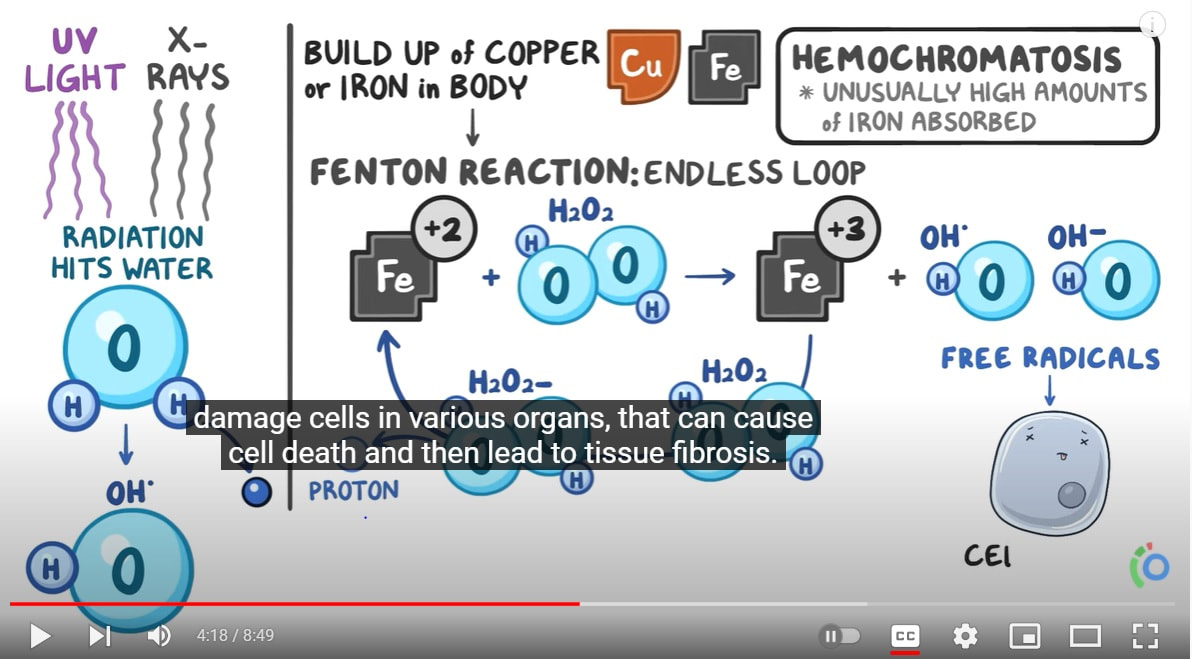
A build up of Copper or Iron in the blood can
04:15: damage cells in various organs, that can cause cell death and then lead to tissue fibrosis.
|
|
Do you suffer from any of these conditions?
Chronic fatigue, fibromyalgia, Lyme disease, Hashimoto’s, rheumatoid arthritis, multiple sclerosis, polycystic ovary syndrome…
The RCP is all about repairing cellular dysfunction to resolve various autoimmune conditions including, but not limited to, the list above. Even though conditions and symptoms will vary from person to person, the fundamental root cause of how they started is the exact same for everyone.
|
Paragraph. ここをクリックして編集する.
|
Paragraph. ここをクリックして編集する.
|
B9
|
Folate - Insufficient cobalamin (B12) slows regeneration of tetrahydrofolate and traps folate in a form that is not usable by the body. This can often be corrected with higher doses of folate but can mask a vitamin B12 deficiency, so vitamin B12 is almost always given when folate is supplemented.
If you decide to supplement with folate, avoid synthetic folic acid. Instead, you should take a biologically active form of folate (methylfolate). Some people have a genetic mutations in the enzyme that produces l-methylfolate in the body; folic acid is a waste and can actually cause harm if you have this genetic mutation. Methotrexate (MTX), widely used in the treatment of rheumatoid arthritis (RA), inhibits dihydrofolate reductase (DHFR) and folate-dependent enzymes. Thymidylate synthase (TS) and methylenetetrahydrofolate reductase (MTHFR) are key enzymes in the folate metabolism and both have been shown to be polymorphic affecting the enzyme activity.(1) |
B1 |
Vitamin B1 is one of the eight water-soluble B vitamins. it plays an essential role in the production of energy from food, the conduction of nerve impulses and synthesis of nucleic acids.
|
B2 |
The main functions of vitamin B2 (riboflavin) are connected to its role as a helper the body to convert vitamin B6 and vitamin B9 into active forms, neutralize ‘free radicals’ that can damage cells and produce energy converting food into glucose. As a result, a deficiency can affect the entire body, leading to low energy, weight gain, and skin and thyroid problems. Lower levels of vitamin B2 have been found in people with depression, so giving them psychiatric medications can actually make them feel worse in the long run.
|
B3
|
Vitamin B3 is one of the water-soluble B vitamins. It is also known as niacin (nicotinic acid) and plays an important role in the disease risk reduction of diseases like Cancer and Diabetes. B3 is depleted by Antidepressant medication.
|
B5 |
Health Benefits of Vitamin B5 include cholesterol and triglycerides reduction in the blood, the acceleration of wound healing -especially following surgery- and help with symptoms of rheumatoid arthritis.
|
B6
|
Vitamin B6 is a key nutrient that boosts mood, deepens sleep, and supports your entire nervous system. Vitamin B6 is commonly referred to as pyridoxine. It is responsible for creating serotonin and norepinephrine in the brain and plays a key role in synthesizing antibodies and forming red blood cells.
|
B9 |
Folic acid (Vitamin B9) is essential for the proper functioning of the body and healthy living. It plays an important role in maintaining healthy digestive system, hair, skin, kidneys and eyes.
|
B17 |
The daily recommended intake for an adult is 30 micrograms (µg), but many biotin supplements marketed for beauty reasons contain much higher doses, ranging from 5,000 µg to 10,000 µg. And some new studies even suggest that mega doses of biotin (100,000 µg to 300,000 µg) could be used to treat diseases, such as neurodegenerative disorders like multiple sclerosis. Most of the published research on biotin interference covers hormone tests, such as parathyroid hormone (PTH), thyroid stimulating hormone (TSH), T4 and T3 tests, as well as tests for troponin. However, because biotin is used in so many immunoassays, scientists say it could interfere with many others. Excess biotin in patients' blood samples can interfere with types of tests called immunoassays because many use biotin as part of the testing methodology which is why this information should be revealed when blood is tested..
|
Headache This is a result of disturbances in some part of the body. Treatments for migraines include the entire Vitamin B complex for health of the nerves.
|
The spleen is an organ located between the stomach and diaphragm.
Leukpenia - Abormal of decrease in white blood corpuscles
Leukocytosis - increase in white blood cells Hematocrit - Percentage of red blood cells per volume of blood spenomegaly - Diagnosed when spleen measures more than 13 cm Nodular - Amyloid is found in the walls of the sheathed arteries and in the follicles but not in the red pulp. Causes of splenomegaly
|
Q: What does your spleen do?
|
|
|
Blood Toxicity
(peripheral blood-based evaluation) |
|
Rheumatoid arthritis patients have an increased risk developing blood clots deep in the veins condition known as venous thromboembolism or VTE which can prove life-threatening if clots break
off and become lodged in the lungs.(1), (2) 
|
Conditions that decrease blood cells from bone marrow:
When you’re sick or have a minor injury, substances that your body sees as foreign, known as antigens, call your immune system into action.
Examples of antigens include:
|
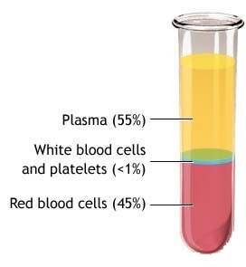
Blood Plasma
A straw colored, sticky fluid which is 90% water and contains over 100 different dissolved solutes such as nutrients, gases, hormones, wastes and products of cell activity, ions and proteins mostly produced by the liver. Synovial fluid is derived largely from your blood plasma. If your blood is toxic, if your blood is deficient in oxygen, if your blood is acidic, it's going to reflect in the synovial fluid.. Toxic Blood and what foods make blood acidic (meat, dairy, coffee, alcohol, wheat, excess sugar) |
Blood Components, Hemoglobin, Type/Rh Factor, Agglutination
|
|
What's Your Blood Type ?
Personalized Nutrition What Your Blood Type Means for Your Health with Dr Peter D’Adamo
James D'Adamo (Hemolologists, Naturopath) believed maybe there's something in the difference in the blood that could explain why people respond differently to these different types of diets. After his death, his son Peter continued his father's work. In 1996 Dr. Peter D'Adamo revolutionized the health and nutrition world with the publication of 'Eat Right For Your Blood Type'. This New York Times bestseller has helped millions of people dramatically improve their health and well-being since then he's written more than 20 books on personalized nutrition.
ABO type, these are called antigens and are found in distribution throughout the human body. Most people are aware that Type O is a blood group that has a lot of protection by virtue of the fact that it carries antibodies to both blood group A and blood group B. If you're blood Type is O you cannot receive blood from a person who the type A or type B, you can only receive from another O.
Antigen: molecules that stimulate a response by T and B cells, surface proteins found on cells or viruses or environmental molecules
( like pollen or asbestos).
Antibodies are proteins found in blood or other bodily fluids of vertebrates, identify and destroy foreign objects such as bacteria and viruses, are proteins that recognize a specific antigen, bind to foreign antigens, which attract macrophages. Abbreviation for antibodies: Ig
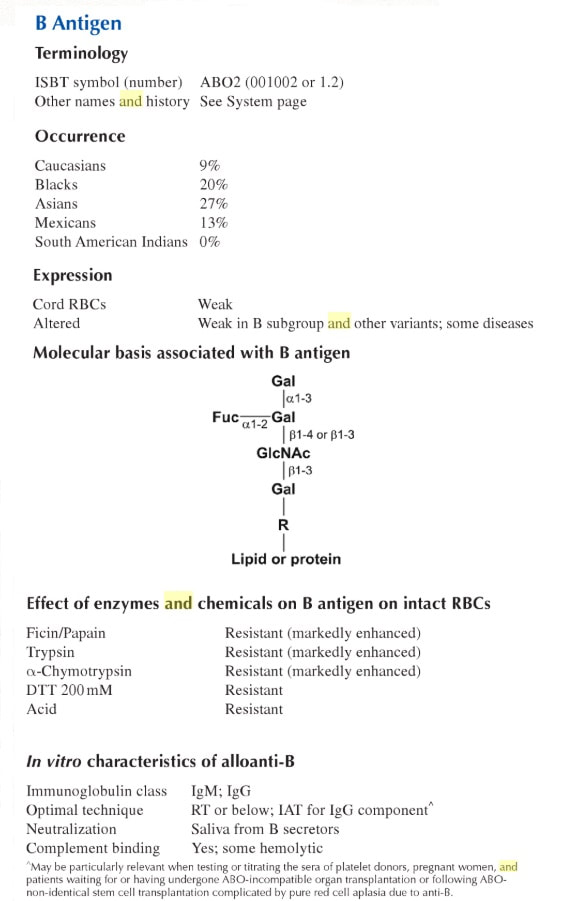
Above image source: The Blood Group Antigen Facts Book
By Marion E. Reid, Christine Lomas-Francis, Martin L. Olsson pg 46-47 
Use of lectins in immunohematologyhttps://www.ncbi.nlm.nih.gov/pmc/articles/PMC4782487/
Anti-B. Lectins showing anti-B activity are relatively less common than those showing anti-A activity. The anti-B specificity was detected in the "old" seeds (more than 1 year since harvested) of Bandeiraea (now Griffonia) simplicifolia, a leguminous shrub from West Africa. 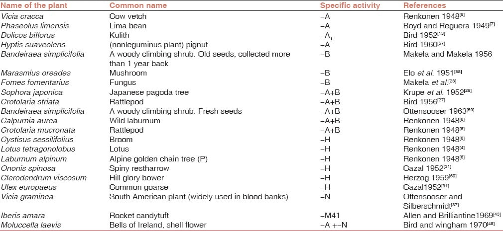
|
Image source: Personalized Nutrition What Your Blood Type Means for Your Health with Dr Peter D’Adamo
The following studies suggest or conclude that lectin may be a contributing pathogenetic factor in RA. (1), (2), (3), (4), (5), (6), (7) ...
There are many types of lectin but one for Blood Group is B, in particular is very interesting. The relationship between exposed galactose and N‐acetylglucosamine on IgG in Rhematoid Arthritis, JCA and SS was investigated. This was achieved using IgG isolated from serum where the levels of galactose and N‐acetylglucosamine (GlcNAc) were detected using biotinylated lectins. Galactose and GlcNAc on IgG from patients with RA and JCA are inversely related, but in contrast, in SS, galactose expression on IgG decreased while GlcNAc expression remained similar to normal levels. Alterations in IgG glycosylation are closely associated with the development of adult and juvenile chronic arthritis and SS, but the changes involved are different in RA compared with SS, suggesting that the precise pattern of exposed sugars is associated with different rheumatological diseases. 1. Modulation of immune function by dietary lectins in rheumatoid arthritis 2. Mannan binding lectin in rheumatoid arthritis. A longitudinal study 3. .Characterization of changes in IgG associated oligosaccharide profiles in rheumatoid arthritis, psoriatic arthritis, and ankylosing spondylitis using fluorophore linked …
The History of the Toxin: Lectin
By the end of the 19th century, it was known that some proteins can agglutinate red blood cells. Earlier, the lectins identified were derived from plants, specifically from seeds of leguminous plants.
Landsteiner who discovered ABO blood groups in 1900 detected hemagglutinating activity in some nontoxic plant extracts. In 1909, he observed that hemagglutinating activity of certain seed extracts was inhibited by either heat-treated serum or by mucin. The fifth decade of the 20th century witnessed a dramatic improvement of research in the area of plant seed extracts. Several scientists published reports of their extensive surveys on the search for seed extracts showing blood group-specific activity. The reports of Renkonen in Helisinki (1948),[6] Boyd and Reguera in Boston (1949),[7] Bird in India (1955),[8] and Boyd in Egypt (1950)[9] were some important reports describing blood group specificity in some seed extracts. Source: https://www.ncbi.nlm.nih.gov/pmc/articles/PMC4782487/ The Beging of the New Theray
Image: List of Dietary Lectins
The Relationship between food and onset of Rheumatoid Arthritis and numerous other diseases. Pending on Blood Type, certain types of foods can react tamiyalogically with you based on your blood type. Watch Video |
Why does Japan care so much about Blood Types?
|
|
|
Furukawa results were presented in a 1926 paper titled “Research on Temperament due to blood type” he came up with these traits for each blood type (above).
Blood Group A
contain anti-B in their serum
Consume:
.did better on a more of a modified Mediterranean / Asian / vegetarian type diet (Plant-based Diet) beans, legumes Eat less of: lima beans, chick peas, navy beans, kidney beans, red meat, - Prone to appendicitis (1)
-type A blood type are negatively impacted by cancer (stomach) and is more commonly attacked by cancers and by bacteria because cancer and bacteria both have a protein that looks like an 'A' and so the body doesn't always fight it. - more resistance to the plague - more susceptible to malaria - more resistant to typhoid - does not make a whole lot of stomach acid - benefit from fermented soy products - problems regulating cortisol - over manufacture cortisol when under stress - stress hormones (and to a lesser degree type B) they go up but they take a long time to come down and that general tendency to have increased cortisol level and cortisol activity is really what does the damage Hawthorn berry is a cardiovascular tonic. It dilates the blood vessels, which helps the blood flow through the arteries and lowers high blood pressure. This beneficial herb for the person with blood type A helps the heart pump more efficiently and may help lower cholesterol levels. |
Blood Group B
contain anti-A in their serum
Consume:
Omnivore diet Turkey, Lamb, soy, potatoes, yams, cabbage Eat less of: Chicken (cells will gluten), corn, buckwheat, soy, rye, certain types of lentils, less sesame, corn oil, sunflower oil, nuts, seeds, tomatoes B blood type are negatively impacted by viruses. The body looks at viruses and notes they have a ' B ' antigen or ' B ' protein and so the body doesn't always fight it once the virus enters, it's hard to get rid of them. 
5-HTP (5-Hydroxytryptophan) is a chemical by-product of the protein building block L-tryptophan. It is sourced from the seeds of an African plant known as Griffonia simplicifolia.
- stress hormones go up but they take a long time to come down and that general tendency to have increased cortisol level and cortisol activity is really what does the damage
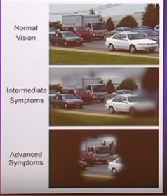
Glaucoma: The Silent Blinding Disease - https://www.youtube.com/watch?v=hRpmnKoAte4
|
Blood Group AB
No antibodies to A or B blood type, If I have antibodies I'll destroy myself
1. Iberis amara
-idiosyncratic diet in the extreme, these people have an A antigen and a B antigen so many foods that react with A or B are sometimes problematic.
- Low stomach acid - gall bladder problems Blood Groups A and B have risk factors for venous thrombosis. (1) |
Blood Group O
Antibodies - contain both anti-A and anti-B in their serum
Consume:
lean meats, oily fish, sea vegetables, green leafy vegetables, did better on a higher protein diet - Paleo diet - buckwheat Eat less of: soy products, wheat, Prone to Conditions:
- autoimune diseases - heart disease - appendicitis (1) - ulcers/erosions (1)- - association for ulcers - chronic Candida infections - susceptible for cholera - prone to depression, bipolar, mental illness due to difficulties maintaining adequate levels of dopamine because they have too much of an enzyme that makes them convert their dopamine to adrenaline.(1)
Blood group O has an enzyme three and a half to four times greater that splits cholesterol, it increases the absorption of calcium (Bone formation through enzymatic hydrolysis of orthophosphate), and has a slight healing effect in the digestive tract in and of itself. (1)
|
Anti T, Anti Tk (peanuts)
Arachis hypogaea Anti T, Anti Tn, Anti Cad (soy bean) Glycine sclarea Anti N
Vicia graminea Bauhinia spp |
American Society of Hematology
1.non-O blood type predicted a higher risk of relapse -rituximab
https://www.hematology.org/search.aspx#?cludoquery=Age%2C%20Blood%20Type%2C%20and%20Rituximab%20Use%20Associated%20With%20Relapse%20Risk%20in%20TTP&cludopage=1&cludorefurl=https%3A%2F%2Fwww.hematology.org%2FPatients%2FTrials.aspx&cludorefpt=Clinical%20Trials&cludorefact=Blood%20type&cludorefaci=1
2. Swedish Researchers Identify Two New Types of Blood Cancer in Children
3. Medicare is covering 10 percent of indications for allogeneic transplant,”(1) Allogeneic stem cell transplantation involves transferring the stem cells from a healthy person (the donor) to your body after high-intensity chemotherapy or radiation.
4.
5.
6.
7.
peripheral blood-based evaluation VS. synovial tissue, the primary site of RA from which tissue and thus disease subtypes are emerging BASED ON synovial tissue.
The treatment of rheumatoid arthritis (RA) has been transformed with the introduction of biologic disease modifying anti-rheumatic drugs (bDMARD) and more recently, targeted synthetic DMARD (tsDMARD) therapies in the form of janus-kinase inhibitors. Nevertheless, response to these agents varies such that a trial and error approach is adopted; leading to poor patient quality of life, and long-term outcomes. There is thus an urgent need to identify effective biomarkers to guide treatment selection. Significant investment in biomarker studies has failed to deliver clinically meaningful tools, with the vast majority focusing on peripheral blood-based evaluation (Lylia Ouboussad ET AL., 2019)
The synovium is the principal target of inflammation in RA, undergoing marked pathological changes compared to healthy tissue. The study of RA synovial tissue has offered insights at a cellular level into multiple aspects of the disease, from identifying pathogenic processes and pathways (3, 4); to explaining clinical manifestations. Furthermore, changes in synovial tissue following successful treatment allow better understanding of mechanism of drug action (5–7).
|
Healthy SynoviumIn health
the synovial membrane contains relatively few cells, consisting of an intimal lining layer of 1–2 cell thickness and a distinct synovial sublining layer (10). The intima comprises fibroblast-like synoviocytes (FLS, also known as synovial fibroblasts or type B synoviocytes) intercalated with macrophage-like synoviocytes (MLS, also called type A synoviocytes) (11). The sub-lining layer is a well-vascularized connective tissue, containing collagen fibers and evenly dispersed FLS and MLS (11). The synovial membrane is key to the structure and function of the healthy synovial joint. The synovial membrane controls transport to and from the synovial cavity, thus maintaining the composition of synovial fluid as well as overall joint homeostasis and integrity. The intimal lining is particularly important, as its lack of tight junctions or a true basement membrane allows the ingress and egress of various cells and proteins (12). Intimal FLS orchestrate proceedings, controlling the synovial fluid volume, secreting hyaluronan for lubrication, clearing intra-articular debris, regulating various immunological processes, and maintaining the extracellular matrix (ECM) of the sublining (13). |
RA SynoviumIn RA
the synovial tissue becomes markedly expanded, with a striking increase in cellular infiltration. This leads to hallmark “pannus” formation at cartilage-bone interfaces; pannus can be composed of macrophages, FLS, leucocytes, plasma cells, and mast cells (14), and behaves like a locally invasive tumor, mediating damage and erosion formation in later disease (15). The intimal lining can expand to 10–20 cells in thickness, partly due to an increase in FLS, but mostly due to infiltration by bone marrow-derived MLS recruited from the circulation (15). Highly activated macrophages send pro-inflammatory signals to intimal FLS, inducing invasiveness, and to B cells, which in turn produce various pro-inflammatory mediators. Paracrine and autocrine signaling networks develop in this way, further propagating synovitis (16). Sub-lining MLS have been associated with disease activity (17) and synovial inflammation measured on magnetic resonance imaging (MRI) (18), and therefore appear of paramount importance to the inflammatory joint reaction (19). Proliferation of FLS are a prime cause of synovial hyperplasia, and major mediators of damage to cartilage and bone, via both direct and indirect interactions, including production of inflammatory mediators, adhesion molecules, proteolytic enzymes and pro-osteoclastogenic factors (13). T cells are able to establish important crosstalk with antibody-producing plasma cells (15, 20, 21). When present, CD3+ T cells in the RA synovium are mostly found in deeper sub-lining layers, where they may be homogeneously or randomly distributed, or clustered in follicle-like structures (19). Similarly, B cells, when present, are mostly organized in follicular structures, which can act as pro-inflammatory, immunological niches (19). |
Personalized RA targeted therapy use.
Synovial Tissue Gene Expression Profiles - (1)
Distinct molecular signatures indicating pathways relating to T cell-mediated immunity and major histocompatibility complex (MHC) class II mediated immunity (amongst others) upregulated in early RA, and pathways relating to the cell cycle upregulated in later disease (40) have been reported. Similarly differential gene expression between high and low inflammatory subsets of RA patients in relation to disease duration has been observed (29).Gene Expression Analysis Across Synovial PathotypesRecently, a machine learning algorithm was able to predict RA synovial gene expression subtype according to 20 histological features. Three subtypes were pre-identified based on RNA-seq clustering: high inflammatory, low inflammatory, and mixed. The high inflammatory subtype showed enrichment of pathways of immunity, immune cell signaling (including SH2, SH3, JAK/STAT, and TNF-mediated signaling), immunoglobulins, chemokines, and cytokines. The low inflammatory subtype was defined by enrichment of transforming growth factor β pathways, glycoprotein synthesis, and cell adhesion genes (45). Distinct myeloid and lymphoid synovial histological subtypes were not identified, in contrast to previous studies (24), but the high inflammatory subtype displayed elevated expression of genes previously attributed to these in the literature.
Synovial Tissue Studies to Predict Response to Biologic and Targeted TherapiesGeneral Synovial Tissue Biomarkers of Response To Therapy
CD68 MacrophageEffective treatment can modify synovial histology, cytokine and gene expression, with ineffective treatment having little impact, thus providing a means to assess for pathological response (46). Synovial sublining (CD68) macrophage numbers and macrophage expressed cytokines have been shown to correlate with disease activity, and change in sublining macrophage to be the optimal indicator of effective therapy, thus providing a potential early predictive biomarker of drug response (6, 47, 48). A recent study demonstrated that the transcriptional profile of isolated RA synovial macrophages highlighted different subpopulations of patients and identified 6 novel transcriptional modules that were associated with disease activity and therapy (49). The authors suggest that transcriptional signatures in macrophages regardless of location (sublining vs. synovial lining) predict responsiveness to specific non-biologic and/or biologic therapies.
Synovial Pathotypes and ResponseA study by Dennis et al. suggested myeloid and lymphoid pathoypes may predict therapeutic sucess with TNF inhibitors (TNFi) and IL-6-targeted tocilizumab, respectively (24). Analysis of serum chemokines further suggested these two pathotypes correlate with raised serum suloble intercellular adhesion molecule 1 (sICAM) and CXCL13 (sICAM/CXCL13) compared to high CXCL13/sICAM, respectively. These initial observations however have not been validated in other cohorts using the serum correlates (50) indicating the need for additional such synovial tissue studies. Nevertheless, stratifying patients by synovial pathotype may inform choice of targeted therapy.
Multiple types of therapies will be discussed in detail below, these are summarized in Table 1 together with key findings which indicate response to biologic and synthetic targeted DMARDs. Table 1. Rheumatoid synovial tissue studies of biologic and targeted synthetic DMARDs.
Anti-cytokine Therapies - Tumor-Necrosis Factor-InhibitorsCell Mediated Therapies -B-Cell Depletion: RituximabSmall Molecule Janus-Kinase (JAK) Inhibitors
|
IgM - first antibody produced when infection occurs
After a B cell has responded to a pathogen's antigen with the generic IgM, future activation will cause the antibodies to "switch" their constant region for better efficiency in a particular environment (blood, mucous membranes, etc.) most appropriate for where that antibody will act in the body. IgA - secretions (breast milk, saliva, sweat, etc) IgD - found on surface of B cells and activate them IgG - babies receive all the mom's various IgG in breast milk, providing immunity to whatever the mom is immune to. IgG crosses the placenta, and is the most abundant (80%,) antibodies in body IgE - allergic reactions Serum sickness
Serum sickness in humans is a reaction to proteins in antiserum derived from a non-human animal source, occurring 5–10 days after exposure. It is a type of hypersensitivity, specifically immune complex hypersensitivity. 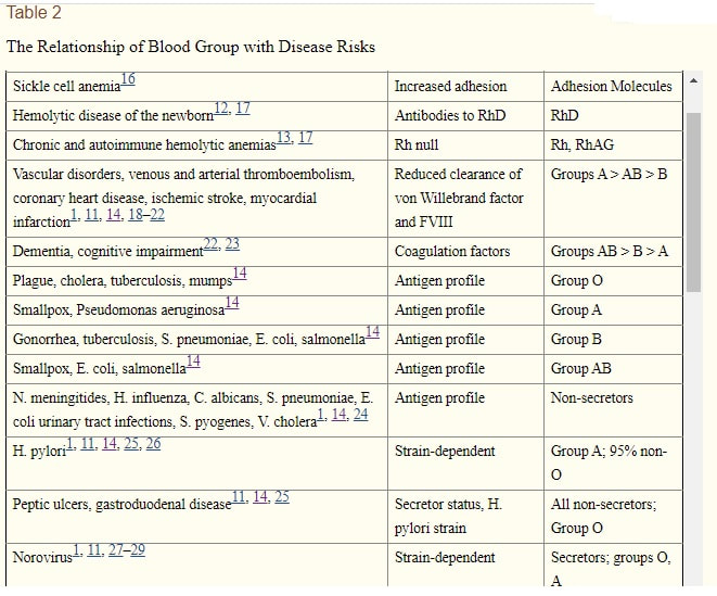
|
Periodontal disease has been associated with a higher risk of having RA. The postulated mechanism is citrullination produced by gingival bacteria.(1)
Al-Katma MK, et al. Control of periodontal infection reduces the severity of active rheumatoid arthritis. J. Clin. Rheumatol. 13(3), 134–137 (2007). 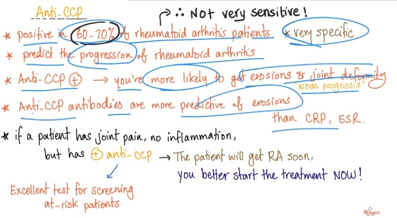
Anti-Cyclic Citrullinated Peptide (anti-CCP)
|
|
Whether as bacterial proteins related to bacterial pathogenicity, or as eukaryotic components of biochemical signalling systems, DING proteins require further study.
Plant DING proteins and their microbial relatives may elicit allergic responses leading to arthritic disease (1). Disease states in which DING proteins have been implicated include rheumatoid arthritis, lithiasis, atherosclerosis, some tumours and tumour-associated cachexia, and bacterial and viral adherence (2). The first DING protein was identified from human rheumatoid arthritis (RA) synovial fluid as a lymphocyte stimulatory protein [3]. DING proteins have also been reported from other animals, e.g., turkey (as a lipid-free polysaccharide-binding protein) and rat (as a cotinine receptor). DING proteins from several plant and fungal species are characterized by short N-terminal sequences and one has been shown capable of binding a germin-like protein in tobacco [5]. Together these studies suggested that DING proteins are widespread in eukaryotes and play important roles in cell signaling and biomineralization. |
Synovial Tissue Heterogeneity in Rheumatoid Arthritis and Changes With Biologic and Targeted Synthetic Therapies to Inform Stratified Therapy
|
A reaction called the glutenation can occur if you certain types of foods that react negatively with your blood type based upon the fact that the food contains a natural protein that acts as an a gluten. Lectins are very very choosing molecules they will interact often with these glycoproteins of the blood group in such a way so that they will have a specificity for one blood type and a different blood type they would leave completely alone.
|
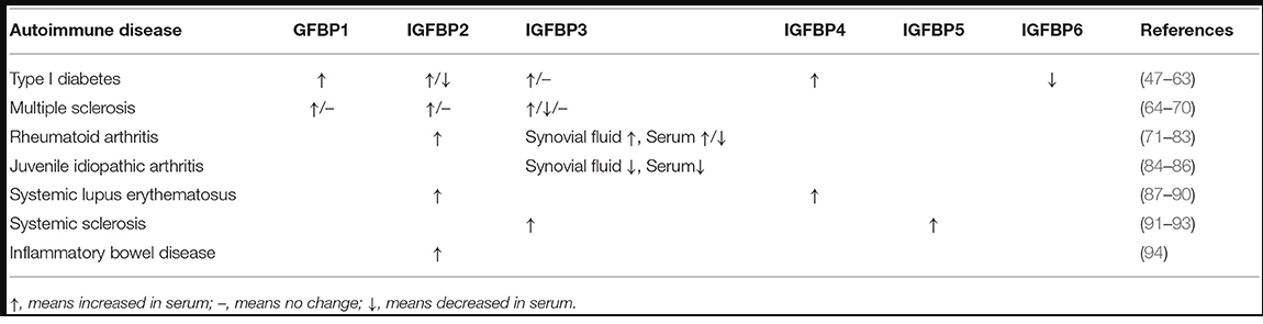
IGFBPs as biomarkers in autoimmune diseases
Despite the advances of researches on IGFBPs as biomarkers in cancer, there have been several studies focusing on the utility of IGFBPs as biomarkers in autoimmune diseases (Table 1), most of which were investigating the diagnostic role of IGFBPs in the disease. While the potential role of IGFBPs as diagnostic biomarkers has been summarized below, the mechanism of action for these molecules has not been widely investigated. Here we summarized studies investigating the potential roles of IGFBPs in the diagnosis and monitor of autoimmune diseases (1).
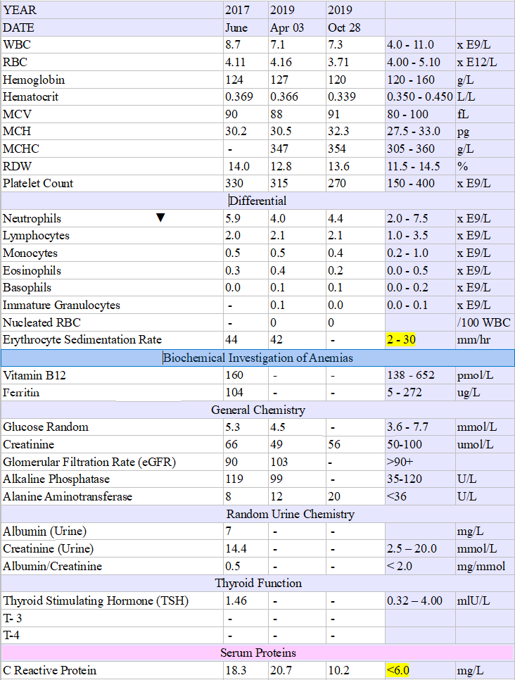
Blood Work: Among the information that can be determined by performing a complete blood count (CBC) are:
The major causes of kidney dysfunction is heart problems: https://www.youtube.com/watch?v=W0OmgjNRSIE
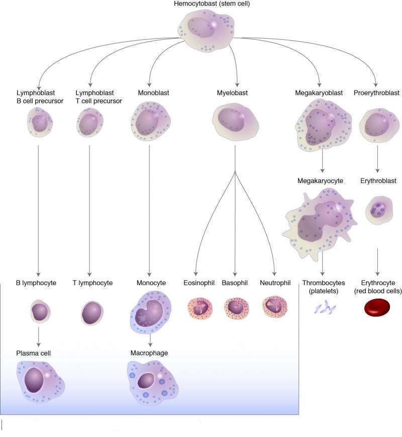
Eosinophil
Eosinophils, sometimes called eosinophiles or, less commonly, acidophils, are a variety of white blood cells and one of the immune system components responsible for combating multicellular parasites and certain infections in vertebrates. Along with mast cells and basophils, they also control mechanisms associated with allergy and asthma. Wikipedia
|
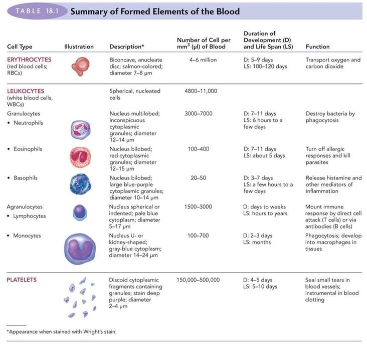
Stem cells in the bone marrow differentiate and mature into one of several types
White Blood Cells (WBCs) There are five different types of white blood cells:
White blood cell count (WBC): An elevated white blood cell count suggests the possibility of an active infection. Patients taking corticosteroids may have an elevated WBC due to the medication.
The white cells have a memory and after having a a virus like chickenpox, the white cells remember that virus and don't allow us to to get that again as long as we have we have an intact immune system, as we get past the age of 70, our testosterone level and growth hormones decrease a lot; those are two hormones that stimulated our immune system and made our immune system reactive. When these levels decrease the white cells lose their memory and we get shingles. That's why some immune immunizations don't work after 70 unless you've been replacing your hormones and stem cells. If you replace your testosterone in both sexes if you stimulate your growth hormone or some people take growth hormones then those two things keep your immune system going. Cells of the Immune System (PART I - GRANULOCYTES) (FL-Immuno/02)www.youtube.com/watch?v=UO6f_EBCZZQ |
Complete Blood Count (CBC)
Hemoglobin (Hgb), the part of each RBC that carries iron
Hematocrit (Hct), the percent of RBCs in the blood
Red blood cell count (RBC): Chronic inflammation can cause a low red blood cell count.
Hemoglobin (Hgb), the part of each RBC that carries iron
Hematocrit (Hct), the percent of RBCs in the blood
Red blood cell count (RBC): Chronic inflammation can cause a low red blood cell count.
- RBC Carry oxygen to and carbon dioxide away from the cells in your body. In Anemia, blood has abnormally low oxygen carrying capacity; it is a symptom rather than a disease itself; blood oxygen levels cannot support normal metabolism; signs/symptoms include fatigue, paleness, shortness of breath, and chills.
|
hematocrit decreased in
|
hematocrit elevated in
|
- The platelet count is often high in rheumatoid arthritis patients, while some potent arthritis medications can cause platelets to be low.
|
VACCINATIONS and IMMUNITY
Dr Nathan Thompson runs laboratory diagnostic work before/after jab 1 and jab 2 and here are the results. www.facebook.com/1177455092/videos/643747873678004 
|
Differential Tests
Erythrocyte sedimentation rate (ESR)
|
|
CCP Anti-CCP RF |
Protein and Antibody Tests
Each of these tests is performed on a blood sample, which may be collected at the same time as the vial(s) taken for your CBC: Anti-cyclic citrullinated peptide (anti-CCP) is an antibody present in most rheumatoid arthritis patients. ... A positive anti-CCP test result can be used in conjunction with other blood tests, imaging tests, and/or physical examination findings to diagnose rheumatoid arthritis.
|
|
|
The D antigen.
Although there are lots of other Rh antigens RH-D is the most significant because it's the most likely of the Rh antigens to produce an immune response. Depending on whether the RH-D antigen is present, each blood type is assigned a positive or negative symbol. People who are Rh-D negative can only receive Rh-D negative blood. But people who are Rh D+ can receive either Rh D positive or Rh D negative blood. The negative blood types, A negative, B negative, AB negative, and O negative are more rare than their positive counterparts. And while the D antigen is the most important one in the Rh system there are a total of 60 other Rh antigens making it the largest of any of the blood classifications. |
Imaging studies are also used to help formulate a diagnosis. Your doctor may order X-rays, which can reveal deformities and abnormalities of bones and joints. These studies are usually ordered initially to help diagnose osteoarthritis.
While useful in this way, X-rays do not show cartilage, muscles, and ligaments. In addition, what is seen on an image doesn't always correlate with what you're experiencing. For example, you may have a lot of pain, though your X-ray doesn't indicate considerable damage—or vice versa.6
Magnetic resonance imaging (MRI) scans produce cross-sectional images of your body by using a magnetic field and radio waves. It can provide precise information about bones, joints, and soft tissues, and detect very small changes in the body.
C-reactive protein level
|
Homocysteine: if a person has B12 or folate deficiency, or if someone has suffered a heart attack or stroke, often their doctor will order a homocysteine test. It’s also a marker for heart disease and can give a bigger picture to the usual blood pressure and basic metabolic panel. Normal levels are 4 – 14 µmol/L and elevated levels are connected to increased risk of heart disease and stroke. |
Rheumatoid factor positive test
|
Rheumatoid factor (RF): antibodies produced by the immune system that can attack healthy joints and tissues.
The presence of RF is not diagnostic or conclusive because RF can be present in hepatitis, SLE, TB, infectious mono, and symphilis (clinical observations and other tests are necessary to confirm RA). Of all rheumatoid arthritis (RA) patients, 65% were positive for rheumatoid factor (RF), and 72% were positive for the shared epitope (SE) (1). |
anti-cyclic citrullinated protein (ACPA) |
anti-cyclic citrullinated protein: autoantibodies (antibodies to an individual's own proteins) that are directed against peptides and proteins that are citrullinated. They are present in the majority of patients with rheumatoid arthritis.
Anti-cyclic citrullinated peptide antibody in rheumatoid arthritis:relation with bone erosion - (LIVER TOXICITY, FRUIT) - When you have seropositive RA, it means your blood has antibodies that can attack your body and inflame your joints. They're called anti-cyclic citrullinated peptides (your doctor may call them anti-CCPs), or anti-citrullinated protein antibodies (ACPAs).Your doctor can give you a blood test to see if you have anti-CCPs. But having them doesn't always mean you have RA. |
Gamma globulin level |
a2- macroglobulin level
|
BNP (Brain Natriuretic Peptid)
|
BNP (Brain Natriuretic Peptid) is a substance secreted from the ventricle chambers of the heart in response to
changes in pressure that occur when heart failure develops and worsens. Level of BNP in the blood decreases when the heart failure condition is stable,
BNP helps physicians make decisions about hospitalizations, aggressive treatments, and future prognosis |
Uric acid
Joint fluid analysis can provide a doctor with many details about the health of a person's joint.5 For certain types of systemic rheumatic diseases, biopsies of certain organs can provide important diagnostic information.
- High levels of uric acid in the blood (known as hyperuricemia) can cause crystals to form which are deposited in the joints and tissues. Deposition of uric acid crystals can cause painful gout attacks. Uric acid is the final product of purine metabolism in humans.
Joint fluid analysis can provide a doctor with many details about the health of a person's joint.5 For certain types of systemic rheumatic diseases, biopsies of certain organs can provide important diagnostic information.
The list below includes some of the autoantibody tests that are used to identify systemic autoimmune disorders. These disorders may cause signs and symptoms associated with inflammation throughout the body.
These are examples of autoantibodies associated with certain systems or organs:
Clotting (coagulation) system
- Antinuclear Antibodies (ANA)
- Antineutrophil Cytoplasmic Antibodies (ANCA)
- Anti-Double Stranded DNA (anti-dsDNA)
- Anticentromere Antibodies (ACA)
- Antihistone Antibodies
- Cyclic Citrullinated Peptide Antibodies (CCP)
- Extractable Nuclear Antigen Antibodies (e.g., anti-SS-A (Ro) and anti-SS-B (La), anti-RNP, anti-Jo-1, anti-Sm, Scl-70)
- Rheumatoid Factor (RF)
These are examples of autoantibodies associated with certain systems or organs:
Clotting (coagulation) system
- Cardiolipin Antibodies
- Beta-2 Glycoprotein 1 Antibodies
- Antiphospholipid Antibodies (APA)
- Lupus anticoagulants (LA)
- Anti-Tissue Transglutaminase (anti-tTG) and Anti-Gliadin Antibodies (AGA)
- Intrinsic Factor Antibodies
- Parietal Cell Antibodies
- Thyroid Autoantibodies (e.g., anti-TPO, TSH receptor antibodies)
- Smooth Muscle Antibodies (SMA) and F-actin Antibody
- Antimitochondrial Antibodies (AMA) and AMA M2
- Liver Kidney Microsome Type 1 Antibodies (anti-LKM-1)
- Anti-Glomerular Basement Membrane (GBM)
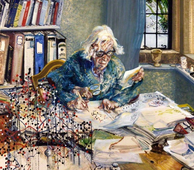
Painting of Dorothy Hodgkin
by Maggi Hambling, 1985 National Portrait Gallery |
Here are the foods which contain the highest levels of this vitamin:
|
|
|
|
|
Notes:
|
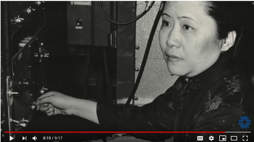
The spin of the cobalt nuclei
03:52
Wu was an impressive scientist. 03:54 She was the best in the business when it came to studying the spin of atomic nuclei. 03:59 So what she decided to do was to set up an experiment in which Cobalt 60 would decay 04:04 via the weak force into Nickel 60, an electron, and an electron antimatter neutrino. ...Video |
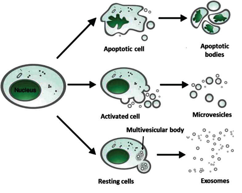
RA may also affect other parts of the body, bringing serious complications such as low red blood cell count (RBC) and inflammation around the heart. Between 0.5 and 1% of adults in the developed world are affected by rheumatoid arthritis, and the numbers are certain to grow as the population ages.(1) New research on neutrophils has revealed that they play a role in protecting cartilage by sending microvesicles, fragments of the plasma membrane, to prevent damage through a complex process involving the proresolving protein annexin A1 and its receptor. The authors envision that microvesicles, with their uncommon knack for reaching cartilage, can be engineered to carry therapeutics, such as omega-3 fatty acids.[1][2]
Open access peer-reviewed chapter
Microvesicles (MVs) Released from Human Red Blood Cells: Properties and Potential Applications
By Duc Bach Nguyen, Thi Bich Thuy Ly and Ingolf Bernhardt
Submitted: October 30th 2016 Reviewed: May 5th 2017 Published: July 12th 2017
|
Although MVs have been discovered for years, the understanding of the mechanism of the formation as well as the biological roles of MVs is still a matter of debate. Recent reported findings led to advances of our understanding of the mechanism of formation and the role of MVs in many different diseases such as vascular diseases, cancer, infectious diseases, diabetes mellitus, diabetes, inflammation, and pathogen infection [24]. Inhibition of the production of MPs may serve as a novel therapeutic strategy for some diseases, especially for cancer treatment [11, 23, 26, 27]. In the next part of this chapter, the biogenesis, properties, and biological function of MVs released from human red blood cells (RBCs) are mainly addressed.
Neutrophil microvesicles protect cartilage in arthritis ... https://www.nature.com/articles/nrrheum.2015.175 Headland, S. E. et al. Neutrophil-derived microvesicles enter cartilage and protect the joint in inflammatory arthritis. |
|
RHEUMATOID ARTHRITIS Neutrophil-derived microvesicles ...https://stm.sciencemag.org/content/scitransmed/7/315/315ra190.full.pdf
RHEUMATOID ARTHRITIS Neutrophil-derived microvesicles enter cartilage and protect the joint in inflammatory arthritis Sarah E. Headland,1 Hefin R. Jones,1 Lucy V. Norling,1 Andrew Kim,2 Patricia R. Souza,1 Elisa Corsiero,1 Cristiane D. Gil,3 Alessandra Nerviani,1 Francesco Dell'Accio,1,4 Costantino Pitzalis,1,4 Sonia M. Oliani,3 Lily Y. Jan,2 Mauro Perretti1* ... |
Neutrophil-derived microvesicles enter cartilage and ...https://stm.sciencemag.org/content/7/315/315ra190.full
Neutrophils play an active role in protecting cartilage from damage by dispatching microvesicles (MVs) to do their bidding in this tissue they otherwise can't access. Headland and colleagues found... |
Neutrophil-derived microvesicles enter cartilage and protect the joint in inflammatory arthritis ...https://www.researchgate.net/publication/284716680_Neutrophil-derived_microvesicles_enter_cartilage_and_protect_the_joint_in_inflammatory_arthritis
In vitro, exogenous neutrophil-derived AnxA1 (+) MVs activated anabolic gene expression in chondrocytes, leading to extracellular matrix accumulation and cartilage protection through the reduction... |
