INFECTION
|
The hidden threat of hepatitis B

|
www.sciencedirect.com/science/article/pii/S2214109X16301449#!
The Lancet Global Health. Volume 4, Issue 8, August 2016, Page e502 Editorial Author links open overlay panelThe Lancet Global HealthAvailable online 19 July 2016, Version of Record 19 July 2016. Tenofovir, then, is a key drug in both the prevention and treatment of chronic hepatitis B in resource-limited settings, yet doubts over its affordability remain. Nayagam and colleagues' costing analysis assumed a generic price of US$48 per person per year, yet currently only HIV programmes are eligible for this reduced price. Additionally, in many middle-income countries such as China, where the mother-to-child transmission study was done, tenofovir is not yet available in generic form: in China it costs a punishing $2920 per person per year. WHO's new strategy calls for 90% of people with chronic hepatitis B to know their status and for treatment coverage to reach 80% of eligible patients by 2030. One could be forgiven for feeling sceptical. But by emulating the HIV story, not least in terms of education, political lobbying, and determined action on drug and diagnostic costs, and by taking advantage of the extensive infrastructure and systems already set up for HIV screening, elimination ought not to be seen as utopian. Hepatitis B is a hidden threat both to public health and to patients themselves, but the tools exist to prevent and treat it. It's time to put them to use. |
Hepatitis A and E viruses
Hepatitis E virus (HEV) in Cuba
G Lemos, S Jameel, S Panda, L Rivera… - Journal of clinical …, 2000 - Elsevier
HEV from its association to hepatitis B or hepatitis C infection, or due to the development of … Cuba. Our findings suggest an important number of the sporadic viral hepatitis cases in Cuba …www.sciencedirect.com/science/
Hepatitis E virus genotype 1, Cuba
MCM Villalba, LAR Lay, V Chandra… - Emerging infectious …, 2008 - ncbi.nlm.nih.gov
… (Ig) M against hepatitis A and hepatitis C viruses. One patient had positive results for hepatitis B surface antigen but negative results for anti-hepatitis B core antigen IgM and hepatitis B …
Dual infection with hepatitis A and E viruses in outbreaks and in sporadic clinical cases: Cuba 1998–2003
LA Rodriguez Lay, A Quintana… - Journal of medical …, 2008 - Wiley Online Library
… From 1998 to 2003, 258 serum samples were collected during 33 outbreaks of acute viral hepatitis from all regions of Cuba (West, Central, and East). Eighteen of these outbreaks took …
A Brief History of Homeoprophylaxis in Cuba, 2004–2014
G Bracho, I Golden - Homœopathic Links, 2016 - thieme-connect.com
… [10] [11] It is also a fact that RCTs of vaccines are not always undertaken; for example, there are no RCTs of hepatitis B vaccine for newborn infants, and some of the multivalent vaccines …
Save Cite Cited by 6 Related articles
Paragraph. ここをクリックして編集する.
Paragraph. ここをクリックして編集する.
Coronaviruses post-SARS: update on replication and pathogenesis.
Coronaviruses post-SARS: update on replication and pathogenesis | Nature Reviews Microbiology
2009 NATURE REVIEWS MICROBIOLOGY Stanley Perlman , Jason Netland
University of IowaParasitologyCoronavirus
Although coronaviruses were first identified nearly 60 years ago, they only received notoriety in 2003 when one of their members was identified as the aetiological agent of severe acute respiratory syndrome. Previously these viruses were known to be important agents of respiratory and enteric infections of domestic and companion animals and to cause approximately 15% of all cases of the common cold. This Review focuses on recent advances in our understanding of the mechanisms of coronavirus replication, interactions with the host immune response and disease pathogenesis. It also highlights the recent identification of numerous novel coronaviruses and the propensity of this virus family to cross species barriers... View Full Abstract Coronaviruses post-SARS: update on replication and pathogenesis. | Paper | Microsoft Academic
Although the severe disease forming capabilities of human coronaviruses were only recognized because of the SARS epidemic, it was well known that animal coronaviruses could cause life-threatening disease. TGEV, which causes diarrhoea in piglets, infectious bronchitis virus (IBV), a cause of severe upper respiratory tract and kidney disease in chickens, and bovine coronavirus (BCoV), which causes respiratory tract disease and diarrhoea in cattle ('winter dysentery' and 'shipping fever'), are all economically important pathogens. Feline infectious peritonitis virus (FIPV), a virulent feline coronavirus (FCoV), causes an invariably fatal systemic disease in domestic cats and other felines. Unlike most strains of FCoV, which are endemic causes of mild diarrhoea, FIPV arises sporadically, most likely by mutation or deletion in felines persistently infected with enteric strains of FCoV70,
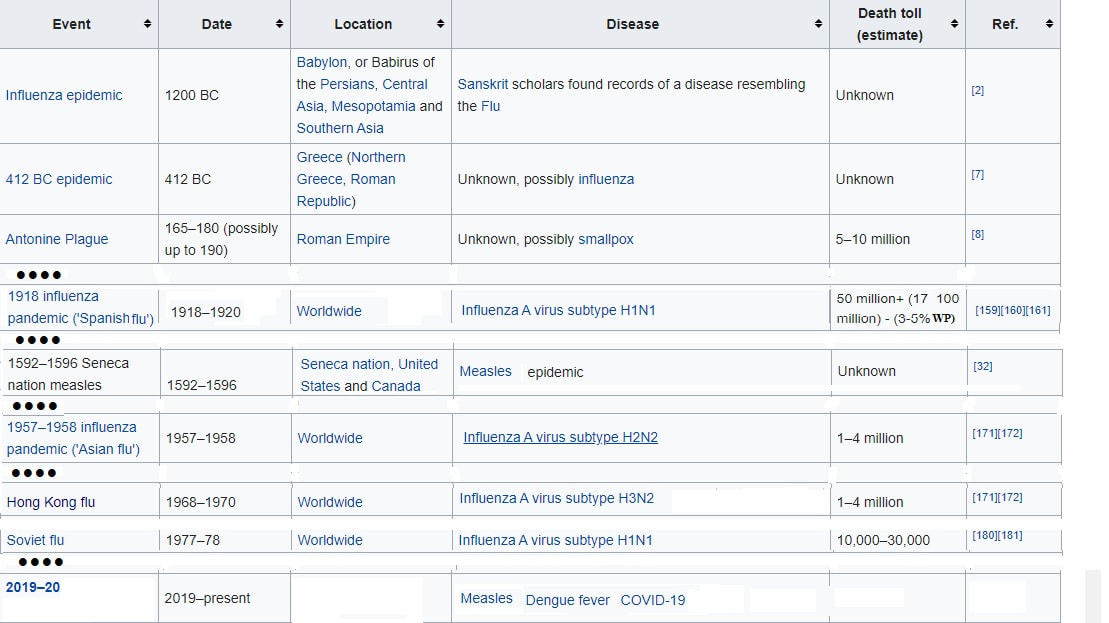
List of epidemics (https://en.wikipedia.org/wiki/List_of_epidemics)
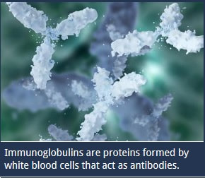
The IgE antibody, as well as IgA and IgG, exclusively function in the immune system and
have distinct and specific jobs to protect the body against damaging foreign cells.
have distinct and specific jobs to protect the body against damaging foreign cells.
|
Antigen: molecules that stimulate a response by T and B cells, surface proteins found on cells or viruses or environmental molecules
( like pollen or asbestos). |
Antibodies are proteins found in blood or other bodily fluids of vertebrates, identify and destroy foreign objects such as bacteria and viruses, are proteins that recognize a specific antigen, bind to foreign antigens, which attract macrophages. Abbreviation for antibodies: Ig
|
Herpes Simplex Virus (HSV) gE/gI Expressed in Epithelial Cells Interferes with Cell-to-Cell Spread
journals.asm.org/doi/full/10.1128/JVI.77.4.2686-2695.2003
Like gD, gE/gI appears to be able to interact with cellular components of cell junctions, gE/gI receptors which can promote HSV cell-to-cell spread.
journals.asm.org/doi/full/10.1128/JVI.77.4.2686-2695.2003
Like gD, gE/gI appears to be able to interact with cellular components of cell junctions, gE/gI receptors which can promote HSV cell-to-cell spread.
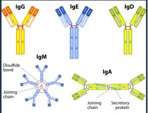
IgA
|
IgM |
IgG
|
IgD
|
IgE
|
|
- 10-15% of antibodies in serum
- Common form in muscous membranes and body secretions (mucus, saliva, tears, breast milk) - In serum, it is a monomer, but forms a dimer consisting of two IgA monomers known as secretory IgA - Enters and passes through a mucosal cell and picks up a polypeptide called secretory component -Secretory component: protects IgA from enzymatic degradation - IgA immunity is relatively short - lived - Prevents the attachment of microbial pathogens to mucosal surfaces - The most common ailment associated with selective IgA deficiency is a greater susceptibility to common illnesses such as ear infections, bronchitis and sinusitis. |
- 5-10% of antibodies in the serum (0.25% of the total amount of immunoglobulin found in the blood serum.)
- Shape keeps it from moving into tissues, generally stay in blood vessels - Appears first in response to a primary function
- IgM is present on younger B cells and is replaced by IgD as the B cell matures. - Many humans have undetectable levels of this antibody, but this does not appear to affect overall immune function. COVID-19 IgM Detection by ELISA Antibody › When IgM is detected you may still be infected or you may have recently recovered ... A negative test result means that the antibodies to the virus that causes .. .. - Macro |
- Gamma Globulin of blood fraction
- 70 - 80% of circulating antibodies - Easily cross blood vessels and enter tissue fluids - Maternal antigen can cross placenta and give passive immunity to the fetus -protect against circulating bacteria viruses neutralize bacterial toxins, trigger the complement system - Parvovirus B19 IgG, also known as fifth disease, is estimated to be contracted at some point by about half of all adults, or possibly more. It typically has mild, flu-like symptoms or no symptoms at all, and it runs its course in a matter of a few weeks. If you are suffering from chronic anemia or other types of iron-deficiency related conditions, your doctor might request the parvovirus B19 IgG test to rule out the possibility that your body is suffering from this infection and cannot fight it on its own. |
- 0-2% of total serum antibodies
- IgD is used to instruct other immune cells to begin producing antimicrobials. -Found in blood, lymph, also on surfaces of B cells -Serum function is unknown but may eliminate B cells that can produce antibodies against self. - uncommon antibody that is produced by immature B cells |
- 0.0002% of total serum antibodies
- This IgE response is very potent and can begin within two to 30 minutes of exposure to the allergen. Release of histamine or other proteins can irritate mucus membranes, cause blood vessels to dilate, or cause smooth muscles of the airways to constrict. Symptoms for the patient can range from sneezing and hay fever to the life-threatening signs of anaphylactic shock. - Bind tightly by Fc(stem) regions to receptors on mast cells and basophils - Mast cells and basophils partcipate in allergic reactions However they can be protective by attracting complement and phagocytic cells against parasitic worms |
IgD antibody with the same function as immunoglobulin M (IgM). Together, these two molecules are responsible for activating B cells when antigens are introduced into the body. Once the B cells are active, they attempt to destroy the antigen by producing other types of immunoglobulins.
IgD is also used to instruct other immune cells, including mast cells and basophils, to begin producing antimicrobials. These compounds help protect the respiratory system from foreign bodies that can enter through the lungs. Scientists believe that there are other purposes for the antibody IgD, though the nature of these other functions is still unknown.
Immunoglobulin E (IgE) is a protein called an antibody that is produced by cells in the body known as lymphocytes. IgE is primarily involved in the allergic response. Some patients can react to various allergens, such as pollen, medications, or food. When a patient encounters an allergen, IgE binds to cells known as mast cells. These mast cells are activated to release granules of histamine, which then produces symptoms of an allergic response.Lymphocytes that originate and mature in the bone marrow are known as B-lymphocytes. These B-cells secrete different types of antibodies or immunoglobulins and are part of the humoral immune response. When a patient is first exposed to an allergen, some of these B-cells are activated and begin to secrete immunoglobulin E. This IgE binds to the surface of mast cells present in tissues of the body. It can also bind to other cells in the blood called basophils.After the first exposure to allergen, the mast cells and basophils are sensitized, but no allergic reaction has taken place. At the time of the second exposure, the allergen binds to the immunoglobulin E molecules on the cell surface. The IgE molecules then become linked to each other by the allergen, which signals the cells to release granules containing histamine, enzymes, or other proteins known as cytokines.
|
Immunoglobulin A, IgA, is produced in the mucous membranes of the body. It is found in all mucous secretions, such as tears and saliva, as well as in the blood. Type A is unique in that it doesn’t break down when exposed to enzymes. This makes it an important disease fighting component in the body. Aside from Type A, there are four other types of immunoglobulin present in the blood. IgG is present in the greatest amount. IgA and IgM are also present in large amounts. The antibodies IgD and IgE are also present, but in much lower quantities.(1)
The mucous membranes of the body are expansive, and, without their protection, would leave an open pathway into the body. A person that suffers from selective Immunoglobulin A deficiency will
have normal levels of the other antibodies in their body. The patient
will also have fully functioning T-cells, phagocytes, and other
components of the immune system. The exclusive absence of Type A
Immunoglobin is what gives the disease the name selective IgA
deficiency.
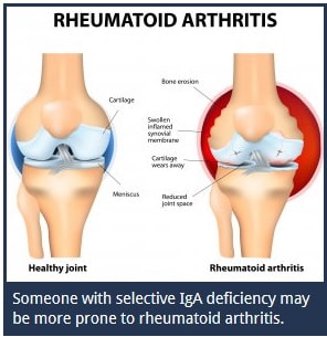
Selective Immunoglobulin A deficiency is a relatively common immunodeficiency illness. People suffering from this ailment may appear perfectly healthy, or they may suffer from problems related to the deficiency. The most common ailment associated with selective IgA deficiency is a greater susceptibility to common illnesses such as ear infections, bronchitis and sinusitis. People that suffer from selective Immunoglobulin A deficiency are more susceptible to autoimmune disorders such as Rheumatoid Arthritis and Lupus. Patients who suffer from a selective IgA deficiency are also more prone to allergies and asthma. Eczema is another condition that occurs in people suffering from this deficiency. |
There are various health conditions that can lead to low levels of Immunoglobulin A in the body. When the body does not have sufficient quantities of Type A Immunoglobin, the person may be diagnosed with selective IgA deficiency. This is considered a disease of the autoimmune system. Some diseases, such as gonorrhea, can destroy Immunoglobulin A in the body, leading to a deficiency.
|
Occasionally, deposits of Immunoglobulin A can build up on the kidneys.
This leads to a condition known as IgA nephropathy. IgA nephropathy is
considered a chronic condition. Doctors are not sure what leads to the
buildup of Type A Immunoglobulin on the kidneys with this ailment.
Low IgA
IgA Test
|
Antigens that induce antibody production
|
Cyclic Citrullinated Peptide
|
Post-Translational Modification of Proteins
|
Anti-Cyclic Citrullinated Peptide (anti-CCP)
|
|
Citrullination or deimination is the conversion of the amino acid arginine in a protein into the amino acid citrulline. Citrulline is not one of the 20 standard amino acids encoded by DNA in the genetic code.
|
Instead, it is the result of a post-translational modification. Citrullination is distinct from the formation of the free amino acid citrulline as part of the urea cycle or as a byproduct of enzymes of the nitric oxide synthase family.
04:02 glyco should tell you that that's sugars |
The immune system can attack citrullinated proteins, leading to autoimmune diseases such as rheumatoid arthritis (RA) and multiple sclerosis (MS).
|
The Diagnostic Value of Antibodies Against Citrullinated or Carbamylated Fibrinogen in Rheumatoid Arthritis
Clin Lab. 2020 May 1;66(5).
Zhenni Wang, Luan Xue, Jun Xie, Naishuo Zhu
BACKGROUND:This study aimed to evaluate the overall diagnostic value of citrullinated or carbamylated fibrinogen antibodies in patients with rheumatoid arthritis (RA).
METHODS:Serum samples collected from 114 patients with established RA, 143 patients with non-RA diseases, and 200 healthy controls were tested by ELISA for citrullinated fibrinogen (Cit-fib), carbamylated fibrinogen (Ca-fib), and chimeric fibrinogen a/b chain citrullinated peptides (CFABCP). Diagnostic indexes and correlations with titers were calculated, cross reactivities of Cit-fib, Ca-fib, and CFABCP were assessed by competition experiments.
RESULTS:With a cutoff ensuring 98% specificity for RA patients versus healthy controls, the sensitivities of Cit-fib and Ca-fib are 66.67% and 24.6%, respectively, while the sensitivity of CFABCP was 74.56%. Cit-fib, Ca-fib, and CFABCP can inhibit reciprocally in competition experiments. As for non-RA patients, the positive rate of Ca-fib was higher than that of Cit-fib and CFABCP.
CONCLUSIONS:Citrullination and carbamylation of fibrinogen both have a role in RA diagnosing, but citrullination is better. The recombination of peptides, CFABCP, has high specificity and considerable sensitivity for diagnosis for RA patients.
IL - Interleukins
IL1: proinflammatory cytokine, activated by inflammasome caspase cascade
IL2: proliferation and differentiation of T cells, secreted by TH1s.
Source is Th1 & Tc. Target is Tc. Action is cause cloning of Tc
IL3: promotes eosinophil production in imflamm response, along with IL9 it'll recruit mast cells, induces macro differentiation in BM
IL4: class switch to IgE, perpetuates TH2 response, along with IL13 itll act M2 macros, activates TH2, secreted by TH2, antagonistic against TH1. Source is Th2. Target is B cell. Action is cause B cell to become P cell to create Antibodies
IL5: promotes eosinophil production in imflamm response, principal cytokine for eosino growth (imp in chronic allergies), secreted by TH2
IL6: along with TGFbeta, it'll activate TH17, pro-inflammatory cytokine
IL7: do T cell development (defective in X linked SCID)
IL9: along with IL3 it'll recruit mast cells
IL10: differentiation into plasma cells (produced by T fh), suppression of inflammation, secreted by TH2, B cell proliferation and differentiation, important for tissue repair and fibrosis, antagonistic against IL12, inhibit T cell activation and MHCII expression
IL12: activates NKCs, activates TH1 along with IFN-gamma, favors maturation of TH1, inflammation thru M1 macros. Source is APC (dendritic cell), Target is Th. Action is cause Th to become Th1
IL13: produced by TH2, epithelial turnover, mucus production, increased contractility of smooth muscle to expel worm, along with IL4 to activate M2 macros, secreted by TH2
IL15: work for NK cell development (defective in X linked SCID)
IL17: Main cytokine produced by TH17, stim fibroblasts, endo cells, and epi cells to produce IL1, IL6, TNFalpha, G-CSF and CXCL8 for inflammation, along with IL22 stim keratinocytes to produce AMPs, implicated in Autoimmune Diseasesof chronic inflammation, stim myeloid and stromal cells to produce G-CSF for neutro production in BM, act stromal cell to make chemokines to attract neutros. Th17 cells, by virtue of their production of IL-17 and IL-17F, are generally thought to be pro-inflammatory and play an important role in host defense against infection, by recruiting neutrophils and macrophages to infected tissues.
IL21: B cell proliferation and survival (plays role in TD B cell activation), differentiation into plasma cells (produced by T fh)
IL22: secreted by TH17s, along with IL17 help to produce AMPs, increases epi cell turnover
IL23: produced by DCs that support TH17 growth and differentiation, inflammation thru M1 macros
IFN-alpha: Source is Th1
IFN-alpha: Target is Macrophages
IFN-alpha: Action is cause increase in phagocytosis
TNF: Source is macrophages & T cells
- Target is body tissues
- Action is cause increase in inflammation & increase in apoptosis
Antibody Titer
Antibody titer is a different concept from antibody dilution. It is used in the diagnosis of infectious and other diseases.
Antibody titer indicates the level of the antibodies in a blood sample, defined as the greatest dilution (lowest concentration) of the blood sample at which an antibody assay, such as ELISA, still produces a detectable positive result. The higher the antibody concentration in the blood, the greater the dilution that will produce a detectable signal. The actual titer value for an antibody will also vary based on the antibody being test, the method used, and the actual laboratory performing the test.
Anti-nuclear/anti-ANA antibodyAntinuclear antibodies (ANA) describes a group of antibodies that react to various components of cell nuclei. ANA testing is used for the diagnosis of several auto-immune diseases, such as systemic lupus erythematosus. ANA tests are performed through indirect immunofluorescence microscopy or ELISA. The indirect immunofluorescence assay is usually performed on the HEp-2 human tumor cell line. Titers of 1:80 or higher are considered positive in the HEp-2 assays [7]. If the immunofluorescence assay is instead performed on rodent tissue substrate, titers of 1:20 or 1:40 or higher have been considered positive [7].
Hepatitis B antibodySeveral antibodies against the Hepatitis B virus (HBV) are measured for the diagnosis and prognosis of acute and chronic hepatitis. They include antibodies against HBV surface antigen (anti-HBs), antibodies against HBV core antigen (anti-HBc), and an antibody against HBV e antigen (anti-HBe). Comprehensive resources are available about these applications from the CDC. Commonly the detection of these antibodies is done through standardized, and often automatic, assay systems, such as Abbott Architect Anti-HBc II for anti-HBc antibodies [8] or Bio-Rad MonoLISA Anti-HBs kit [9].
References
- Das N. Commercializing High-Concentration mAbs. BioPharm International. 2016;29(11):2016.
- Chew H, De Lima P, Gonzalez Cruz J, Banushi B, Echejoh G, Hu L, et al. Endocytosis Inhibition in Humans to Improve Responses to ADCC-Mediating Antibodies. Cell. 2020;180:895-914.e27 pubmed publisher
- Taube J, Aktürk G, Angelo M, Engle E, Gnjatic S, Greenbaum S, et al. The Society for Immunotherapy in Cancer statement on best practices for multiplex immunohistochemistry (IHC) and immunofluorescence (IF) staining and validation. J Immunother Cancer. 2020;8: pubmed publisher
- Jung H, Fattet L, Tsai J, Kajimoto T, Chang Q, Newton A, et al. Apical-basal polarity inhibits epithelial-mesenchymal transition and tumour metastasis by PAR-complex-mediated SNAI1 degradation. Nat Cell Biol. 2019;21:359-371 pubmed publisher
- Collino C, Jaldin Fincati J, Chiabrando G. Statistical criteria to establish optimal antibody dilution in flow cytometry analysis. Cytometry B Clin Cytom. 2007;72:223-6 pubmed
- Smith A. Finding the best antibody dilution by repeated immunostaining of the same tissue section. Biotech Histochem. 2014;89:215-9 pubmed publisher
- Kavanaugh A, Tomar R, Reveille J, Solomon D, Homburger H. Guidelines for clinical use of the antinuclear antibody test and tests for specific autoantibodies to nuclear antigens. American College of Pathologists. Arch Pathol Lab Med. 2000;124:71-81 pubmed
- Ismail A, Devakumar S, Anantharam R, Fletcher G, Subramani T, John G, et al. Low frequency of occult hepatitis B infection in anti-HBc seropositive blood donors: experience from a tertiary care centre in South India. Blood Transfus. 2012;10:230-2 pubmed publisher
- Behzad Behbahani A, Mafi Nejad A, Tabei S, Lankarani K, Torab A, Moaddeb A. Anti-HBc & HBV-DNA detection in blood donors negative for hepatitis B virus surface antigen in reducing risk of transfusion associated HBV infection. Indian J Med Res. 2006;123:37-42 pubmed
A few supporting vitamins of the immune system include vitamin A, a number of B vitamins, and vitamin C. Vitamins A and C are crucial for immune support, as are the B vitamins.
|
White and red blood cells are produced in the spongy tissue called bone marrow. This substance, rich in nutrients, is crucial for properly functioning immunity. Leukemia, a cancer of the bone marrow, causes greatly increased production of abnormal white blood cells and allows immature red blood cells to be released into the body. Other features, such as the lowly nose hair and mucus lining in the lungs, help trap bacteria before it gets into the bloodstream to cause an infection.
|
|
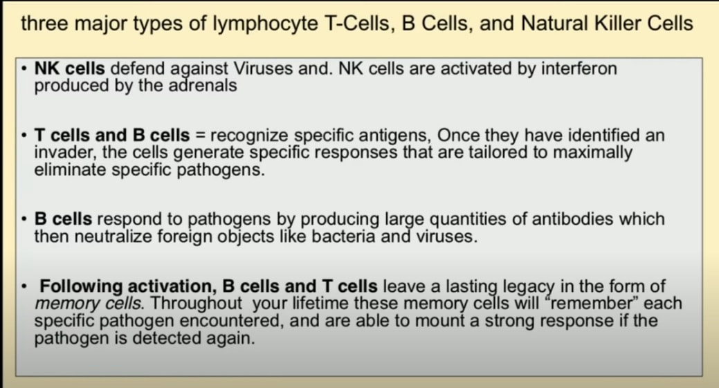
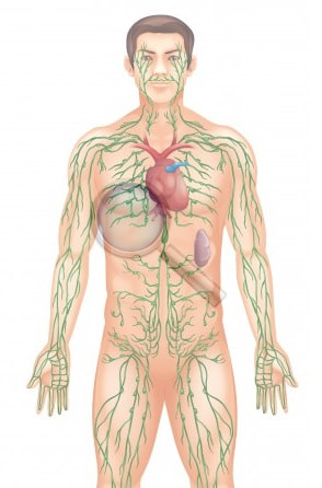
The main component of the immune system is the lymphatic system. Small organs called lymph nodes help carry lymph fluid throughout the body. These nodes are located most prominently in the throat, armpit and groin. Lymph fluid contains lymphocytes and other white blood cells and circulates throughout the body.
The white blood cells are the main fighting soldiers in the body's immune system. They destroy foreign or diseased cells in an effort to clear them from the body. This is why a raised white blood cell count is often an indication of infection. The worse the infection, the more white blood cells the body sends out to fight it. B cells and T cells
are the main kinds of lymphocytes that attack foreign cells. B cells
produce antibodies tailored to different cells at the command of the T
cells, the regulators of the body's immune response. T cells also
destroy diseased cells.
|
Effective Treatment of Severe COVID-19 Patients with Tocilizumab
Xiaoling Xu, (et al. #These authors contributed equally to this work) 1Respiratory and Critical Care Medicine, The First Affiliated Hospital of University of Science and Technology of China (Anhui Provincial Hospital), Hefei, Anhui, People’s Republic of China.
Study demonstrated that in the pathogenesis of SARS, a cytokine storm occurred , involving a considerable release of proinflammatory cytokine including interleukins (IL) -6, tumour necrosis factor α (TNF-α), and IL-12. 11 In the research of Middle East respiratory syndrome, caused by another coronavirus (MERS-CoV), cytokine genes of IL-6, IL-1β, and IL-8 can be markedly high. A delayed proinflammatory cytokine induction by MERS-CoV was also confirmed. 12 Similar to the changes in SARS and MERS, in COVID-19, higher plasma levels of cytokines including IL-6, IL-2, IL-7, IL-10, granulocyte-colony stimulating factor (G-CSF), interferon-γ-inducible protein (IP10), monocyte chemoattractant protein (MCP1), macrophage inflammatory protein 1 alpha (MIP1A), and TNF-α were found in ICU patients, which implied a cytokine storm occurred7,9 and related to the severity and prognosis of the disease. In the biopsy samples at autopsy from a patient who died from the severe infection with COVID-19, histological examination showed bilateral diffuse alveolar damage with cellular fibromyxoid exudates. Mononuclear inflammatory lymphocytes were seen in both lungs. 13 These studies suggested that an inflammatory factor or a cytokine storm have occurred. In our previous research, after analyzing the immune characteristics of patients with COVID-19, we found that aberrant pathogenic T cells and inflammatory monocytes are rapidly activated and then producing a large number of cytokines and inducing an inflammatory storm. Among them, GM-CSF and IL-6 are the key chinaXiv:202003.00026v1 3 cytokines leading to inflammatory storm which may result in increased alveolar-capillary blood-gas exchange dysfunction, especially impaired oxygen diffusion, and eventually lead to pulmonary fibrosis and organ failure. 14 Therefore we suggested that IL-6 might play a key role in the cytokine storm and interfering of IL-6 might be a potentially therapeutic for severe and critical COVID-19. IL-6 receptor has two forms: membrane bound IL-6 receptor (mIL6R) and soluble IL-6 receptor (sIL6R). IL-6 binds to sIL-6R to form a complex, which then binds to gp130 on the cell membrane to complete trans-signal transduction and play a pro-inflammatory role. 15-18 As a recombinant humanized anti-human IL-6 receptor monoclonal antibody, Tocilizumab can specifically bind sIL-6R and mIL-6R and inhibit signal transduction. It is currently used mainly for rheumatoid arthritis. 18 The results of long-term toxicity tests on animals showed that tocilizumab was well tolerated, and no significant abnormalities were observed in other clinicopathological studies or histopathological evaluations. 18-20 In this study, we retrospectively observed tocilizumab in treating severe or critical COVID-19 patients to see if IL-6 plays a pivot role in the parthenogenesis and the efficacy of the tocilizumab interference of IL-6, in order to provide new therapeutic strategy for this fatal disease.
Therefore, tocilizumab can effectively treat severe patients of COVID-19, which might be explained by the blocking of IL-6-associated febrile and inflammatory storm response. Nevertheless, there are several shortcomings in this study. The number of patients were rather limited. It was a single observation study and a significant bias could possibly be existed. Apparently, the evidence strength need to be enhanced. To confirm the conclusions of our observation, a randomized controlled trial and a study on the mechanism of IL-6 in COVID-19 are being under performing.
Important Information
Tocilizumab affects your immune system. You may get infections more easily, even serious or fatal infections. Call your doctor if you have a fever, chills, aches, tiredness, cough, skin sores, diarrhea, weight loss, or burning when you urinate.
Tocilizumab may also cause a perforation (a hole or tear) in your stomach or intestines. Tell your doctor if you have a fever and stomach pain with a change in your bowel habits. Tocilizumab may also cause liver problems. Tell your doctor right away if you have right-sided stomach pain, vomiting, loss of appetite, tiredness, dark urine, clay-colored stools, or yellowing of your skin or eyes.
Tocilizumab may also cause a perforation (a hole or tear) in your stomach or intestines. Tell your doctor if you have a fever and stomach pain with a change in your bowel habits. Tocilizumab may also cause liver problems. Tell your doctor right away if you have right-sided stomach pain, vomiting, loss of appetite, tiredness, dark urine, clay-colored stools, or yellowing of your skin or eyes.
Tocilizumab
|
https://www.drugbank.ca/drugs/DB06273
Interleukin (IL)-6 plays essential roles not only in the immune response, but also in haematopoiesis and the central nervous system. Unregulated production of IL-6 has been found in chronic inflammatory autoimmune diseases, such as rheumatoid arthritis (RA), systemic onset juvenile idiopathic arthritis (soJIA), Crohn's disease (CD), systemic lupus erythematosus (SLE) and vasculitis. Furthermore, IL-6 activities can explain many symptoms of these diseases. More importantly, serum levels of IL-6 are correlated with disease activity. Tocilizumab binds specifically to both soluble and membrane-bound IL-6 receptors (sIL-6R and mIL-6R), and has been shown to inhibit IL-6-mediated signaling through these receptors. ToxicityMost common adverse reactions (incidence of at least 5%): upper respiratory tract infections, nasopharyngitis, headache, hypertension, increased ALT. |
|
Quinine
|
Quinine is a medication used to treat malaria and babesiosis. This includes the treatment of malaria due to Plasmodium falciparum that is resistant to chloroquine when artesunate is not available.
NOTE: Artesunate Artesunate is a medication used to treat malaria. The intravenous form is preferred to quinine for severe malaria. Often it is used as part of combination therapy, such as artesunate plus mefloquine. It is not used for the prevention of malaria What is quinine in tonic water? Tonic water is a soft drink containing quinine, which gives it a bitter taste. Quinine is a common treatment for malaria. Some people believe that it can also help with leg cramps and restless legs syndrome. Quinine comes from the bark of the cinchona tree. Beginning in 2006, the US Food and Drug Administration (FDA) issued a series of warnings not to prescribe the malaria drug quinine (Qualaquin™) for nocturnal leg cramps -- an off-label use -- because it may result in serious and life-threatening hematologic adverse effects.Oct 8, 2012 Quinine and Leg Cramps: Not Worth the Risk - Medscape |
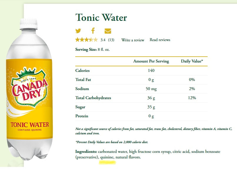
Is there quinine in Schweppes tonic water?
Tonic water contains no more than 83 mg of quinine per liter--a much lower concentration than the 500 to 1,000 mg in the therapeutic dose of quinine tablets. Drinking a few ounces of tonic water shouldn't be harmful, but it isn't likely to prevent your leg cramps. |
|
|
|
|
|
Humans and animals have coexisted since the beginning of time, sharing viruses, bacteria, and perhaps the etiology of cancers. Excluding ionizing radiation, sunlight, and tobacco, infection represents the main known cause of human cancers throughout the world. Read More: www.ncbi.nlm.nih.gov/pmc/articles/PMC3923154/
Of the 217 viruses and prions, 538 bacteria and rickettsia, 307 fungi, 66 protozoa and 287 helminths, known to be pathogenic to humans, 868 (61%) are zoonotic, that is, they can be transmitted between humans and animals. Read More: www.ncbi.nlm.nih.gov/pmc/articles/PMC1088493/ |
The CCR5-Delta 32 mutation has a specific impact on T-cells. 5-13% percent of the population has this gene.
A genetic mutation known as CCR5-delta 32 is responsible for the two types of HIV resistance that exist. CCR5-delta 32 hampers HIV's ability to infiltrate immune cells. The mutation causes the CCR5 co-receptor on the outside of cells to develop smaller than usual and no longer sit outside of the cell.Oct 6, 2013
HIV Resistant Mutation | Viruses101 | Learn Science at Scitable
HIV Resistant Mutation | Viruses101 | Learn Science at Scitable
|
|
Yersinia Pestis:
- causative agent of bubonic plague - causes an intestinal infection in fleas, in mammals it is an intracellular parasite that causes a blood infection centered in and around the lymph nodes - cause of the Black Death in the 14th century A village in Derbyshire, was also badly affected by the Great Plague of 1665 even though the disease is most associated with its impact on London. The sacrifices made by the villages of Eyam may well have saved cities in northern England from the worst of the plague. The village is noted for an outbreak of bubonic plague, which occurred there in 1665, in which the villagers chose to isolate themselves rather than let the infection spread. Eyam Isolated population that survive 50% of population had gene. CCR5-Delta 32 Gene prevented plague from entering the hosts white blood cells. |
Light based anti-infectives: ultraviolet C irradiation, photodynamic therapy, blue light, and beyond
Owing to the worldwide increase in antibiotic resistance, researchers are investigating alternative anti-infective strategies to which it is supposed microorganisms will be unable to develop resistance. Prominent among these strategies, is a group of approaches which rely on light to deliver the killing blow. As is well known, ultraviolet light, particularly UVC (200–280nm), is germicidal, but it has not been much developed as an anti-infective approach until recently, when it was realized that the possible adverse effects to host tissue were relatively minor compared to its high activity in killing pathogens. (1)
BACTERIA, Tooth Decay
32:02
so because I'm a dentist I was very interested in obviously the tooth decay issues. So one of the first things I looked at was what were they doing on tooth decay and in 1950, There are pretty clearly stating their position that the aim of the foundation and dental research has been to discover effective means of controlling tooth decay by methods other than restricting carbohydrate intake, so they were doing things like trying to discover a vaccine for tooth decay creating enzymes that we could maybe put into our toothpaste or even to our foods that would break up the plaque on your teeth so you could keep eating sugar but have a plaque busting toothpaste it didn't work but they're still trying. Source: (1) FoodGate: The Break-in, the Cover-up, & the Aftermath - YouTube https://www.youtube.com/watch?v=-s5szfPYKY4 |
Nematodes
While most of the thousands of nematode species on Earth are not harmful, some cause diseases in humans and other animals or attack and feed on living plants.
https://www.youtube.com/watch?v=EevqmzkGJaE |
The lymphatic system's role in immunity
Learn about how B and T cells reside in lymph nodes. Find out how that enables them to get a preview of what they need to be prepared to fight. By Patrick van Nieuwenhuizen
|

|
Select Agents and Toxins List (https://www.selectagents.gov/compliance/guidance/nucleic/index.htm)
HHS Select Agents and Toxins
Select Agents and Toxins
- Abrin [6]
- Bacillus cereus Biovar anthracis [1]
- Botulinum neurotoxins [1][6]
- Botulinum neurotoxin producing species of Clostridium [1]
- Conotoxins (Short, paralytic alpha conotoxins containing the following amino acid sequence X1CCX2PACGX3X4X5X6CX7) [6]
- Coxiella burnetii
- Crimean-Congo haemorrhagic fever virus
- Diacetoxyscirpenol [6]
- Eastern Equine Encephalitis virus [4][5]
- Ebola virus [1]
- Francisella tularensis [1]
- Lassa fever virus
- Lujo virus
- Marburg virus [1]
- Monkeypox virus [4]
- Reconstructed replication competent forms of the 1918 pandemic influenza virus containing any portion of the coding regions of all eight gene segments (Reconstructed 1918 Influenza virus)
- Ricin [6]
- Rickettsia prowazekii
- SARS-associated coronavirus (SARS-CoV) [5]
- Saxitoxin [6]
- Chapare
- Guanarito
- Junin
- Machupo
- Sabia
- Kyasanur Forest disease virus [5]
- Omsk hemorrhagic fever virus [5]
- Variola major virus (Smallpox virus) [1]
- Variola minor virus (Alastrim) [1]
- Yersinia pestis [1] The most common forms of Plague are: Bubonic Plague, Septicemic Plague, and Pneumonic Plague
- Bacillus anthracis [1]
- Bacillus anthracis Pasteur strain
- Brucella abortus
- Brucella melitensis
- Brucella suis
- Burkholderia mallei [1]
- Burkholderia pseudomallei [1]
- Hendra virus
- Nipah virus
- Rift Valley fever virus
- Venezuelan equine encephalitis virus [4][5]
- African horse sickness virus
- African swine fever virus
- Avian influenza virus [4]
- Classical swine fever virus [5]
- Foot-and-mouth disease virus [1][5]
- Goat pox virus
- Lumpy skin disease virus
- Mycoplasma capricolum [4]
- Mycoplasma mycoides [4]
- Newcastle disease virus [3][4]
- Peste des petits ruminants virus
- Rinderpest virus [1]
- Sheep pox virus
- Swine vesicular disease virus [5]
Select Agents and Toxins
